| Format | What it preserves in an anatomy model | Planning use |
|---|---|---|
| STL | Polygon mesh geometry for a shape to be printed. Check whether colour or separate layers survive your export. | Send to a slicer for 3D printing. Keep a layered master file in an authoring tool. |
| OBJ | Polygon mesh geometry. Accepted on the Kivisense platform alongside GLB, GLTF and FBX. | Move a model between tools when no rig or animation is needed. |
| FBX | Skeletal, transform and blend shape animation. Kivisense caps bone influence at 4 bones per vertex. | Authoring and rigging output. Convert to GLB before web delivery. |
| glTF / GLB | Node hierarchy, materials, cameras, meshes and animations. Textures travel as JPEG or PNG images. | Web viewers and embedded 3D, where small files and fast loading matter. |
The glTF figures follow the fileformat.com glTF documentation, and the FBX and platform limits follow the Kivisense model upload guidelines.
The right choice among 3d anatomy model file formats depends on the destination: GLB for web viewers, FBX for rigged or animated work, and a mesh format such as STL for printing. Match the file to the task before exporting.
Every 3D human anatomy model starts life in an authoring tool and is then exported for a specific use. For the stages before export, see how are 3d anatomy models made.
Choosing a format by task
A format is a delivery decision, not a quality ranking. The same femur or heart can exist as several files, and each file suits a different downstream job.
- Web viewer or embedded 3D: choose GLB. The Kivisense documentation lists GLB, GLTF, FBX and OBJ as supported uploads, yet it strongly recommends GLB. That is the clearest published steer toward GLB for browser delivery.
- Rigged or animated teaching models: choose FBX as the working output. Kivisense recommends exporting FBX from modelling software, then converting it to GLB for upload. Its platform accepts transform, skeletal and blend shape animation, but not vertex animation.
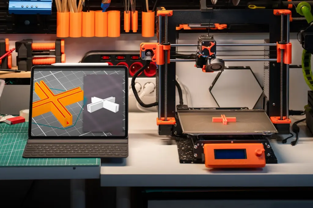
- 3D printing: choose STL. The polygon mesh is what a slicer turns into print instructions. Check your slicer’s import list before assuming another format will load. SciePro says its anatomy models have been tested and validated for professional-grade 3D printing workflows, and the NIH 3D Print Exchange is an open website for biomedical print files, as described in the SIU School of Medicine 3D printing guide.
- Moving a model between programs: choose OBJ when you need plain geometry and no animation. Keep the master scene in the authoring program. The SIU guide notes that Blender supports the whole 3D pipeline: modelling, rigging, animation, simulation and rendering.
Printing and web delivery pull in opposite directions, so the next question is what each format actually carries along with the geometry.
What colour, texture and organ layers survive
Only some formats keep more than raw shape. The glTF description includes node hierarchy, materials, cameras and descriptors for meshes and animations, according to the fileformat.com glTF page. A node hierarchy gives an anatomy model a place to keep organs, bones and vessels as separate, named parts instead of one fused surface.
Textures are stored apart from the geometry. The same documentation describes binary .bin files that hold geometry and animation data, with textures supplied as separate image files. For compatibility with modern web browsers, JPEG and PNG are the supported image formats.
Practical limits tighten at the upload stage. Detailed anatomy art can exceed that.
- For layered study models: build the organ separation in the scene hierarchy before export, and check it survives by opening the exported file.
- For colour: use a format that carries materials and image textures, such as glTF or GLB, rather than assuming a print mesh will keep painted detail.
- For rigged models: keep the FBX or authoring file as the master, because the web file is a delivery copy.
File weight and geometry limits decide whether a model loads at all.
Using FBX and GLB in a web viewer
An FBX human anatomy model is best treated as an authoring file rather than something to load directly in a browser. Kivisense’s advice is to export FBX from modelling software and convert it to GLB for upload, even though its platform technically accepts FBX.

Why GLB suits web viewers
The glTF format stores 3D model information in JSON. The fileformat.com documentation says it was designed for small file size, fast loading, complete 3D scene representation and extensibility. Those goals are what a browser viewer needs, because a student waiting on a large download is less likely to explore the model.
Geometry budgets for the browser
Triangle count is the other limit. It advises modelling in quads, with triangles reserved for special cases. A polygon mesh of a dissection-grade organ can exceed that budget, so the web copy usually needs a lighter version than the print or archive copy.

Web delivery is the end of the pipeline. Many anatomy meshes begin earlier, as medical scans.
From CT scan to printable mesh
Medical imaging data is stored as DICOM images, and free software turns those images into a 3D model. The SIU School of Medicine guide names InVesalius3 and 3D Slicer as free tools for creating 3D models from DICOM scans. The software builds a surface from the scan, and that surface is then saved as a mesh file for printing, with STL the usual print choice.
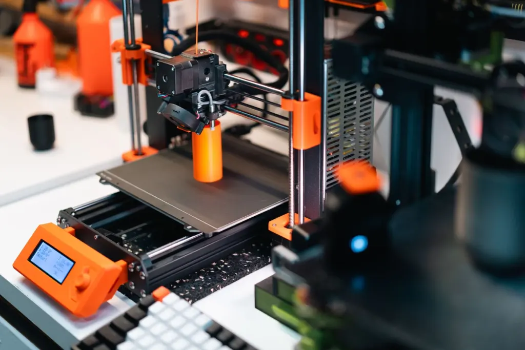
The same guide points to ready-made alternatives when no scan is available. BodyParts3D represents anatomical concepts as 3D structure data for an adult male whole-body model, and the NIH 3D Print Exchange hosts biomedical files built for printing.
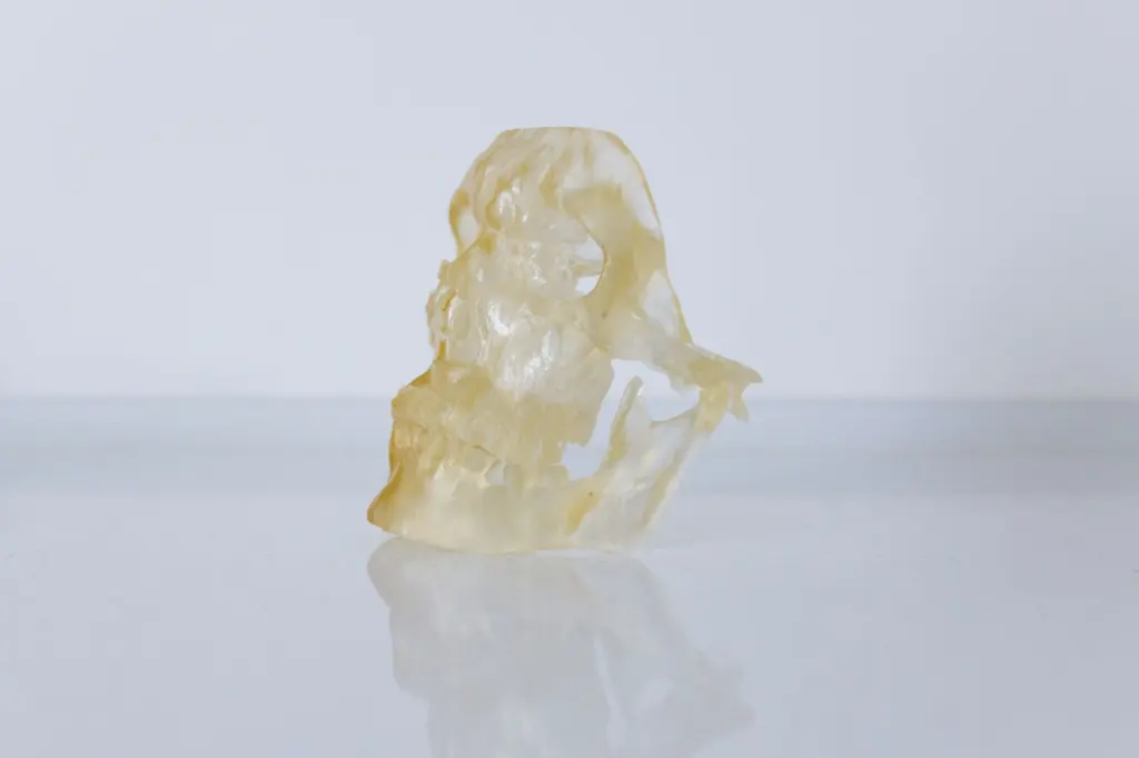
A scan-derived mesh carries shape rather than teaching polish. For adding labels, layers and structured study on top of a finished model, see how to study anatomy with 3d models.
Frequently asked questions
Which file format is best for a 3D anatomy model?
No single format wins. GLB is the strongest choice for web viewers, FBX suits rigged or animated authoring, and a mesh format such as STL suits printing. Keep an authoring master so each delivery copy can be regenerated.
Can glTF or GLB files use Draco compression on every platform?
No. Kivisense states that Draco compression is not supported, and it also rejects meshopt and quantization in glTF or GLB uploads. Check the target viewer’s rules before compressing.
How large should an anatomy model be in a viewer?
Kivisense recommends keeping the model’s size consistent with real-world objects. Its guidelines also cap bones at 4 per vertex and limit textures to 2048×2048 pixels.
When was glTF 2.0 published?
glTF 2.0 was published on June 5, 2017 at the Web3D 2017 Conference, according to the fileformat.com documentation.