A 3D human anatomy model is a digital body, or body region, built from separate named structures that you can rotate, layer and inspect on screen. Start by deciding whether you need to study it, render it or prepare it for printing.
Studying favours interactive platforms with labelled structures. Animation, rendering and print preparation depend more on how cleanly the geometry and textures are built. If budget is the first constraint, begin with the guide to best free 3d human anatomy software, then return here to judge what you are looking at.
What a 3D human anatomy model actually is
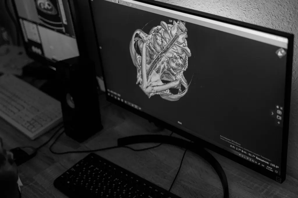
A 3D human anatomy model is a digital reconstruction of the body or a body region, stored as separate geometry for each structure. That separation makes it useful. Software can show a single bone, hide a layer, label an organ or animate a moving part, none of which a flat illustration can do.
BioDigital describes its platform as an interactive 3D software platform for visualizing anatomy, disease and treatments within the human body. It also says its virtual human is made up of over 14,000 individual anatomical structures, which is a vendor claim about scale. The XR Anatomy site sits at the other end of the range, with free interactive 3D models, written descriptions and ways to test your knowledge. Both fit the definition, yet they suit different people.
3D modeling is the construction work behind all of this. A modeller builds a mesh for each structure and then adds the surface data that controls how it looks. SciePro, which supplies professional model libraries, says on its 3D models page that all its models use clean, production-based topology with no unnecessary triangles and no intersecting geometry. Read a statement like that as a quality promise you can test by opening the file, not as a figure that settles quality.
It helps to treat a model as a data product rather than a picture. A picture is judged by looking at it. A data product is judged by what it contains, how it was built, which formats it arrives in and what you are allowed to do with it. The sections below follow that order, starting with the question that decides everything else.
Decide what the model has to do
The job decides which model is worth having, so settle it before comparing libraries. A model that is excellent for revision can be awkward for animation, and a model prepared for production can lack the labels and quizzes that make study efficient.
- Studying on screen. Choose a platform with labelled structures, descriptions and self-testing. BioDigital points to over 200 quizzes in its Human Library, and XR Anatomy pairs models with descriptions and knowledge tests. The approach to using these tools well is covered in how to study anatomy with 3D models.
- Rendering and animation. Choose models that arrive as production assets, with clean topology and texture data you can take into your own scene. SciePro frames its library for people rendering high-end visuals, and the Sketchfab listing for an animated body model shows how animation is packaged. The craft side is covered in 3d human anatomy for animation.
- Immersive learning or apps. Look for models stated to work in real-time engines and extended reality. SciePro says its models are built to perform in those environments, and the Sketchfab listing describes its model as VR/AR ready. For headset and phone use, see augmented reality human anatomy.
- Device and implant design. Geometry matters more than labels here, so a study platform is rarely the right starting point. The specific considerations are set out in 3d anatomy models for implant design.
- Physical output. SciePro states that its models were tested and validated for professional-grade 3D printing workflows through a collaboration with Stratasys. That is a vendor claim, but it names the kind of check to ask any supplier about. The practical side is in 3d printing human anatomy models.
These uses share one test: what can you do with the model after you have looked at it? A browser platform lets you explore and quiz. A production asset lets you move the geometry into other software. Decide which of those you need, and most of the choice is already made.
In education the common case is simple. Students, teachers and self-learners want to explore structures, see relationships and check recall, so interactivity and quizzes outweigh texture resolution. In design and visual work the priorities reverse, and mesh quality, texture maps and delivery format come first.
Body systems and regions in 3D anatomy sets
Anatomy sets are usually organised by body system or by region, and coverage varies a good deal between products. A public Sketchfab listing for an animated human body gives a concrete example of a broad set. Its contents list includes an animated skeleton, a human brain, an animated digestive system, lungs, a heart, the diaphragm, a urinary system and a liver.
That one listing shows the usual pattern. Skeletal structures, the principal organs and a few animated systems are bundled together, and deeper detail often lives in separate specialist sets. Check coverage against your syllabus or brief before you commit, because a set that looks complete can omit the one system you need.
Skeleton
Bones have clear boundaries and are easy to separate. XR Anatomy advertises more than 700 osteology structures in its 3D Osteology App, which indicates how finely a dedicated bone set can be divided. For a deeper look at what a bone set should include, see the 3d human skeleton model guide.

Skull and head
The head is a dense region, and many sets treat it separately from the rest of the skeleton. XR Anatomy, for example, offers anatomy of the cranium with augmented illustrations. If your interest is the cranial bones or head structures, a dedicated 3d human skull model resource will serve you better than a whole-body set.
Muscular system
The muscular system is the layer that matters most when the question is movement. Study sets use muscle layers to show attachments and relationships, while animators use them to understand how surface form changes in motion. The 3d muscle anatomy guide covers that layer. Choose muscle models that sit correctly over the skeleton you are using, because mixing sets from different suppliers can leave structures misaligned.
Internal organs
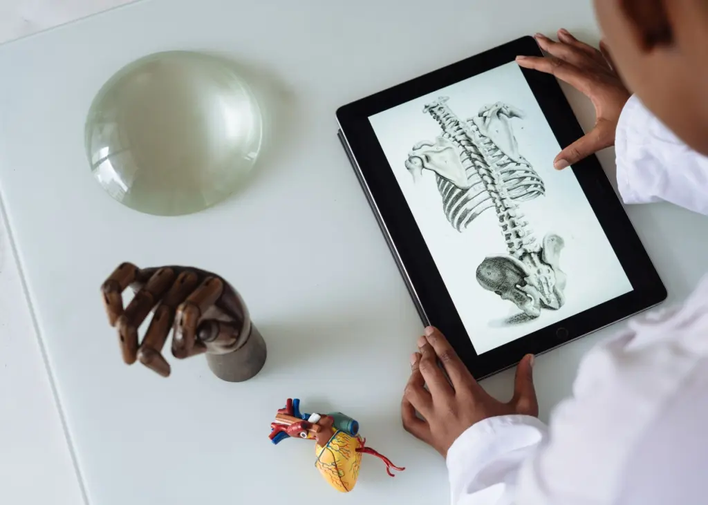
Internal organs are where interactivity pays off, because overlapping structures are hard to follow on paper. The 3d human organs model guide covers whole-torso sets, and the Sketchfab listing above shows organs delivered as individually named parts. The heart gets particular attention from vendors. SciePro describes its heart model as animated with real cardiac dynamics, and XR Anatomy offers interactive 3D heart models. For the detail, see 3d heart anatomy.
One point to keep clear is that a structure count in a vendor’s description measures inventory, not accuracy. SciePro says its SP ONE model includes over 3600 individual structures, and BioDigital cites over 14,000 individual anatomical structures for its virtual human. Those figures count different things in different products, so they cannot be ranked against each other.
Hand and spine
Regional models suit focused study, such as a joint, a limb or a column of vertebrae. The hand is a good example, with many small bones that are hard to see on a whole-body model. The guide to 3d hand anatomy addresses that region, while 3d spine anatomy covers the vertebral column, where being able to rotate and separate individual vertebrae is the main advantage.
Medical imaging and the source data behind models
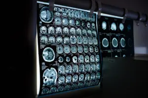
Medical imaging is one of the inputs that can sit behind an anatomical model, but vendors rarely itemise it. BioDigital says its anatomical models are heavily scrutinised and based on a combination of surgical and gross dissection photographs and diagnostic imaging. It does not name a scanning modality or give scan parameters. A model built from a mix of sources is best treated as a composite reference rather than a record of one person’s body.
That distinction matters for the choice you make. For learning normal anatomy, a carefully assembled composite is a sensible reference. For work that must reflect a particular patient or a specific scan, an idealised library model is the wrong tool, and you would need geometry derived from that individual’s imaging instead. If the answer to which scans were used is not on a product page, ask the supplier directly before relying on the model for anything beyond general reference.
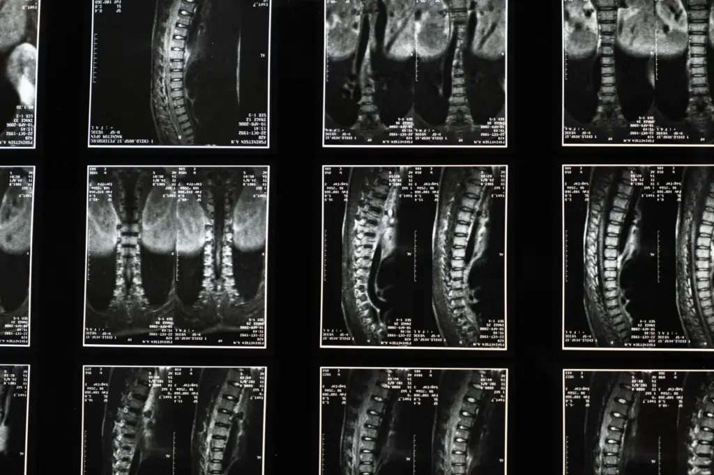
Turning imaging and reference material into finished, named, separated structures is a long editorial process, not a one-click export. The workflow is explained in how are 3d anatomy models made. For now, the practical lesson is that provenance deserves as much attention as polish, because a beautiful render can hide a thin evidence base.
Build quality: topology, textures and mesh size
Build quality is where production models separate from casual ones, and it can be read from a few concrete attributes. Mesh density, topology and texture data tell you whether a model will behave in animation, render cleanly or hold up on the hardware you have.
- Mesh size. The Sketchfab listing for the animated body model reports 222.4k triangles and 111.3k vertices. Those counts give a sense of scale for a whole-body animated model. Compare any model’s counts against the device it must run on, because a mesh that suits a workstation render may be heavy for a phone.
- Topology. SciePro states that its models use clean, production-based topology. The claim has no numeric limits, so verify it by inspecting the mesh for stray triangles and overlapping surfaces before building a project on it.
- Textures and materials. SciePro says its models carry 8K UDIM texture maps, displacement maps and PBR workflows. The Sketchfab listing likewise says its model was created for a PBR approach with metal and roughness values. Material-ready models save time in rendering, but they matter less if you only want to explore structures in a browser.
- Animation. The Sketchfab model includes animated systems, and SciePro animates its heart with cardiac dynamics. Animation is useful for seeing function, but check whether it is baked into the file or driven by the platform.
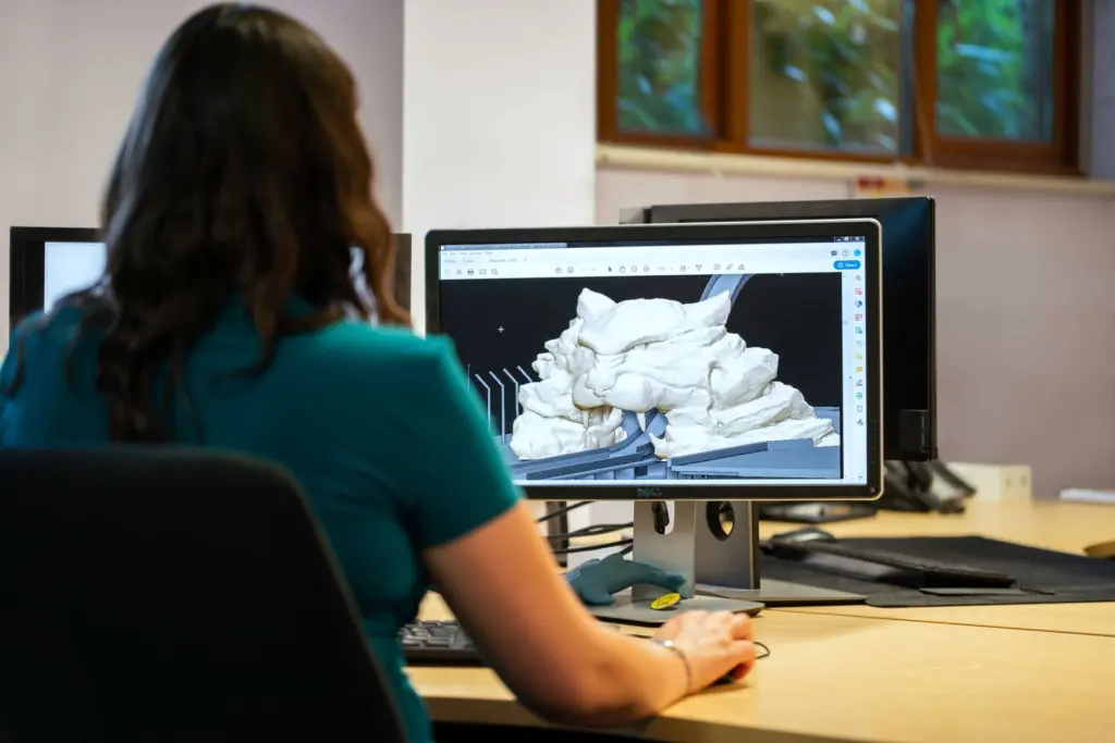
The practical rule is to match the specification to the output. A student browsing structures does not need 8K textures, and an animator cannot work from a model with messy geometry. Paying for or downloading more resolution than the job needs adds weight without adding learning value.
Formats and delivery
How a model is delivered shapes what you can do with it more than most buyers expect. BioDigital says its Human platform uses only HTML standards, unlike solutions that rely on game engines. For a learner, that means exploring anatomy in a browser without installing extra software.
Production libraries work the other way round. SciePro says its models are built to perform in real-time engines and extended reality environments, which implies files you load into your own software rather than a viewer you use inside someone else’s. The Sketchfab listing sits between the two, offering a hosted viewer with model information shown on the page, including mesh counts, before you commit.
When you are comparing options, ask what happens to the model when it leaves the platform. If it stays in a browser viewer, you are buying access. If it comes as files, you are buying an asset, and the file format, texture workflow and licence determine what you can build. The comparison of exports and their trade-offs is in 3d anatomy model file formats.
Digital and physical models compared
Digital models excel at layering, isolation, labelling and testing, while physical models offer a tangible object and need no screen. Neither replaces the other for every purpose, and the best choice depends on what you are trying to learn or produce.
- Layering and isolation. A digital model can hide the structures in front of the one you are studying. A physical model shows what its maker chose to include.
- Self-testing. Platforms such as BioDigital, with over 200 quizzes in its Human Library, turn a model into a recall tool. A physical model offers no built-in testing.
- Handling. A physical model can be held, turned and compared by several people at once without any software. That suits group teaching and quick reference.
- Moving between the two. The categories overlap. XR Anatomy lets users scan printed anatomical illustrations with an iOS device to see structures pop out in augmented reality, and SciePro validates its digital models for professional-grade 3D printing.
For most learners the sensible approach is to treat the digital model as the main reference and bring in a physical model only where handling adds something. For makers, digital comes first by definition, since a printed model begins as a file prepared for print.
Starting points by role
Your role narrows the choice quickly, and a first step is usually clear once you know what the output is.
- Anatomy student. Begin with a free interactive platform that offers descriptions and quizzes, and add regional guides as your syllabus moves from system to system.
- Teacher or course designer. Prefer browser-based delivery, because it removes installation barriers for a class. Check that the structures and systems you teach are all covered before building lessons around a platform.
- Animator or visual artist. Look for production topology, texture maps and animation-ready assets, and inspect the mesh before committing to a project.
- Developer building a VR or AR app. Look for models stated to work in real-time engines and extended reality, and compare mesh counts against your target hardware.
- Designer or maker. Check print validation and provenance, and ask how the model’s source data was assembled.
Whatever the role, keep the decision grounded in the job. Define the output, check the contents against it, inspect the build quality and confirm the delivery format. That order avoids paying for polish you will not use and missing the one attribute that decides whether a model works.
Frequently asked questions
Are there free 3D anatomy models worth using?
Yes. XR Anatomy offers free interactive 3D models with written descriptions and ways to test your knowledge, which is enough for many revision tasks. Free tools are usually narrower than paid libraries, so check that they cover the systems you need.
Can I compare structure counts between vendors?
Not reliably. BioDigital cites over 14,000 structures and SciePro cites over 3600 in its SP ONE model, but the products differ in scope and in how structures are counted. Use counts to gauge ambition, not to rank products.
Do I need a game engine to use a 3D anatomy model?
Not always. BioDigital says its platform uses only HTML standards, so it runs without a game engine. Production libraries such as SciePro’s are aimed at people loading models into real-time engines or extended reality tools.
Can the same model be used for study and for AR?
Sometimes. The Sketchfab animated body listing describes its model as VR/AR ready, and XR Anatomy uses augmented reality to bring printed illustrations to life. Confirm the specific model is stated as suitable for the platform you plan to use.