Quick answer: Learn spinal cord anatomy from the outside in. The cord runs from the base of the brain to about the first and second lumbar vertebrae, shows a butterfly-shaped gray core inside white matter, and continues as the cauda equina.
This guide suits anyone building a mental model of the cord before moving on to detail. For the wider picture of how the cord joins the rest of the central nervous system, start with our overview of brain anatomy.
Choosing a study order for the spinal cord
The cord is easiest to remember when you move from large features to small ones. Each step below answers a different question, and the later steps depend on the earlier ones.
- Start with gross form if you are new to the topic. Knowing where the cord sits and how far it extends makes every later detail easier to place.
- Move to the cross section once the overall shape is clear. The gray and white matter pattern is the same idea repeated along the cord, so it rewards a careful look.
- Study the lower end separately. The conus medullaris and cauda equina confuse many learners because the cord stops well above the bottom of the spine.
- Finish with roots and nerve counts. The pairs of spinal nerves connect the cord to the body, so they make most sense after you know the cord’s levels.
Tract-level detail, meaning which fiber bundle carries which signal, is a later layer. The sections here stay with the cord’s shape and its cross-sectional organization.
Spinal cord anatomy at a glance
The table collects the values most worth memorizing and says how to use each one when you study.
| Feature | Value | Planning use |
|---|---|---|
| Lower end of the cord | Between the first and second lumbar vertebrae in the adult spine | Use this as the anchor level for the conus medullaris and cauda equina. |
| Spinal nerves | 31 pairs emerge between the vertebrae | Treat 31 pairs as the headline count when asked how many spinal nerves there are. |
| Meningeal coverings | 3 layers, as in the brain | Learn the coverings alongside the cord so that its protection is part of the picture. |
| Gray matter shape | Butterfly-shaped center | Use the butterfly as a quick cue when identifying a cross section. |
| Cauda equina | Nerve bundle below the cord end, resembling a horse’s tail | Remember it as roots, not cord tissue. |
These values come from the University of Maryland Medical System spine guide and the Merck Manual overview of the spinal cord.
Gross form and regions of the cord
The spinal cord is a column of millions of nerve fibers that runs through the spinal canal, according to the University of Maryland Medical System. A soft bundle of nerves, it extends from the base of the brain to the lower back, as the Cigna health library describes it.
The cord and the spine are both divided into four regions from top to bottom: cervical, thoracic, lumbar and sacral, as the ECU Health library explains. The regions give you a way to name a level and to link it to what the nerves there supply. For example, nerves of the cervical spine go to the upper chest and arms.
Cord length compared with spine length
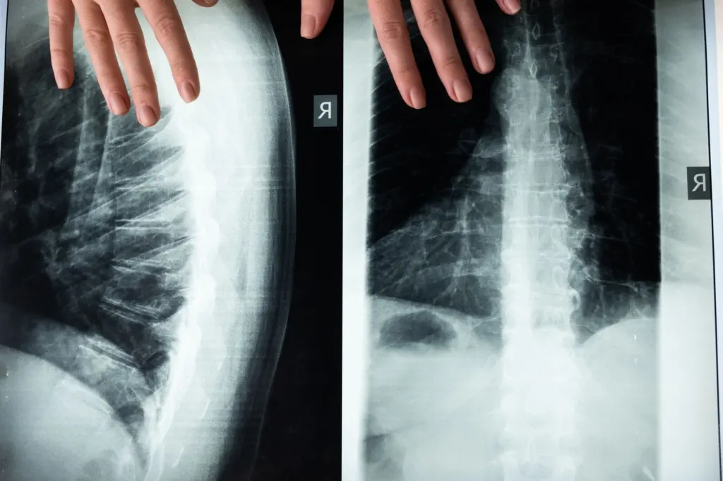
The cord is shorter than the spine. The ECU Health library notes that the lumbar region is where the cord ends, and the Merck Manual places that end about three-fourths of the way down the spine. This mismatch explains why the cord’s levels do not line up neatly with the vertebrae beside them, and why the lower spinal canal holds mostly nerve roots.
Counting vertebrae
Published vertebral counts differ by source. The University of Maryland guide describes 24 small bones, while the Merck Manual says the spine in most adults is composed of 33 individual back bones. If you are revising for a course, follow the count your own textbook uses and keep the cord-level facts separate from it.
Spinal cord cross section: gray matter and white matter
A cross section shows gray matter and white matter arranged in a fixed pattern. According to the Merck Manual, the gray matter forms a butterfly-shaped center in the cord, and the surrounding white matter contains columns of nerve fibers, which are bundles of axons.
What the gray matter contains
The butterfly’s front wings are called the anterior or ventral horns, and they contain motor nerve cells. When you see a cross section, finding the front wings tells you where the motor cell bodies sit. Together with the front-back orientation of the roots, this helps you orient any section you meet.
What the white matter contains
The white matter wraps the gray matter and is organized into columns of axon bundles that run up and down the cord. These bundles are the ascending and descending tracts. For now, treat them as the cord’s conduction lines and the gray matter as the processing center at its core.
Gray matter versus white matter at a glance
- Position: gray matter sits in the center, while white matter surrounds it.
- Shape: gray matter is butterfly-shaped, whereas white matter appears as columns around that shape.
- Content: the anterior horns of the gray matter hold motor nerve cells, and the white matter holds axon bundles.
Coverings and protection
Like the brain, the cord is covered by three layers of tissue called the meninges, as the Merck Manual states. A protective membrane called the dura mater covers the cord, according to the University of Maryland guide, and the ECU Health library adds that the entire cord is surrounded by cerebrospinal fluid, or CSF.
When you sketch a cord in its canal, include these coverings and the fluid. They explain why the cord is described as sitting within protective layers inside the bony vertebral canal rather than touching the bone directly.
Where the spinal cord ends: conus medullaris and cauda equina
The spinal cord ends between the first and second lumbar vertebrae in the adult spine, according to the University of Maryland guide. The terminal end of the cord is the conus medullaris. Below that level the canal no longer contains cord tissue in the usual sense, because the cord continues as a bundle of nerve roots in the lower back, as the ECU Health library describes.
What the cauda equina is
The Merck Manual explains that this bundle is called the cauda equina because it resembles a horse’s tail. The University of Maryland guide adds that the collection of nerves is called the cauda equina while it is still inside the spinal canal. After that, the roots leave through their own openings between the vertebrae.
The cauda equina carries nerve impulses, both motor and sensory, to and from the legs. The ECU Health library notes that the lowest part of the spinal nerve roots contains five pairs of nerves, which is a useful figure when you picture how crowded the lower canal is with roots.
Why this region needs its own study
Many learners assume the cord runs the full length of the spine. It does not, and the difference matters whenever you try to connect a vertebral level with a cord level or a nerve root. Keep two ideas apart: the cord ends where the conus medullaris is, and the roots carry on as the cauda equina.
Roots and spinal nerves
Thirty-one pairs of spinal nerves emerge from the cord between the vertebrae, according to the Merck Manual. The University of Maryland guide describes the same arrangement as the spine branching into thirty-one pairs of nerve roots.
Each nerve attaches to the cord by two roots. The sensory or posterior root is at the back of the cord, and the motor or anterior root is at the front. That front-back split mirrors the motor anterior horn in the cross section, so one mental picture serves for both. For how those roots combine and branch beyond the canal, continue with spinal nerves anatomy.

Answering the segment question
If you are asked how many spinal cord segments there are, the supported figure is 31 pairs of spinal nerves, one for each pair of roots emerging between the vertebrae. Pair that number with the four regional names so you can say both how many and where.
Troubleshooting common mix-ups
- The cord seems to reach the tailbone. It does not. The cord ends near the lumbar region, and the roots below it are the cauda equina.
- A cross section looks unfamiliar. Look for the butterfly-shaped center first, then check which wings face the front, because the anterior horns hold motor nerve cells.
- Vertebral counts disagree between sources. Different guides report different totals for the spine, so use your course’s figure for vertebrae and the 31 pairs for spinal nerves.
- Gray and white matter keep swapping in memory. Remember that white matter surrounds and carries axon bundles, while gray matter sits at the core.
Frequently asked questions
What is the structure of the spinal cord?
The cord is a soft column of millions of nerve fibers inside the spinal canal, from the base of the brain to the lower back. It has a butterfly-shaped gray center, surrounding white matter, and three meningeal layers, as described by the Merck Manual.
What is the difference between gray and white matter in the spinal cord?
Gray matter forms the central butterfly shape and its anterior horns contain motor nerve cells. White matter surrounds it and consists of columns of nerve fiber bundles, according to the Merck Manual.
Where does the spinal cord end?
In the adult spine it ends between the first and second lumbar vertebrae, which the University of Maryland Medical System describes. That is above the bottom of the spine, so the cord is shorter than the spinal column.
What are the conus medullaris and cauda equina?
The conus medullaris is the cord’s terminal end. The cauda equina is the bundle of nerve roots that continues below it, named for its resemblance to a horse’s tail, and it carries motor and sensory impulses to and from the legs.
How many spinal cord segments are there?
The cord gives rise to 31 pairs of spinal nerves that emerge between the vertebrae. The ECU Health library groups the cord into cervical, thoracic, lumbar and sacral regions.