Brain anatomy is easiest to learn by deciding first whether you need the brain’s regions or the wider nervous system. Start with the forebrain, midbrain and hindbrain for the brain itself, or the central and peripheral divisions for the whole system.
This page is the orientation point for the topic. Once you know which route suits you, the detailed guide on parts of the brain and their functions takes the region-by-region list further, and the sections below send you to the right follow-up pages.
Deciding where to start
Your starting point depends on what you are trying to do. The choices below run from the broadest view to the finest scale, so pick the one that matches your task and ignore the rest until you need them.
- You want the big picture of the whole system. Begin with how the nervous system is divided. The central and peripheral split gives every other structure a place to sit, and it is the quickest way to stop confusing the brain, the spinal cord and the nerves that branch through the body.
- You want to name and locate brain regions. Begin with the forebrain, midbrain and hindbrain. This is the route for anyone working with a model, because those regions follow the physical layout you see when you turn a model on its side.
- You want to understand how the brain reaches the body. Begin with the brainstem and spinal cord. They form the physical link, and the nerves of the peripheral system continue from there.
- You want the cell-level view. Begin with neurons, then the difference between grey matter and white matter. This route suits anyone whose course moves from tissue to function rather than from whole organ to part.
- You are studying for a practical or exam. Follow the order your course uses for its models and diagrams, and use this page to fill gaps. Spatial learners usually do better moving from outside to inside, so go from the cerebral cortex to the brainstem, then to the spinal cord.
If you are unsure, start with the nervous system divisions. They take little time and make every later description easier to place.
How the nervous system is divided
The nervous system is organised into two main parts, the central nervous system (CNS) and the peripheral nervous system (PNS), as the Queensland Brain Institute describes it. Everything else in this guide hangs from that split.

Central nervous system
The CNS is made up of the spinal cord and the brain, according to the Dana Foundation. It is the part enclosed in bone. The brain sits inside the skull, and the spinal cord is protected by the bony vertebrae of the backbone. On a model this is easy to see: the brain and cord are a continuous column, and the bony structures are what you remove or cut away to expose it.
Peripheral nervous system
The PNS is the nerves and other types of supporting cells that branch throughout the rest of the body, per the same Dana Foundation overview. The simplest way to hold the difference in mind is by location: the CNS is the brain and spinal cord, while the PNS is everything nervous that lies outside them. If you want a dedicated side-by-side treatment, the guide on central vs peripheral nervous system develops the contrast.
Autonomic nervous system
The autonomic nervous system deals with processes that occur reflexively, such as breathing and heart rate. The Dana Foundation notes that the brainstem is responsible for these autonomic processes, which is why the brainstem receives so much attention in any study of vital functions. To see how the autonomic system is subdivided and what each part does, continue to the comparison of the sympathetic vs parasympathetic nervous system.
With the divisions settled, the brain itself is the next place to look, and it is easiest to take region by region.
The brain region by region
The brain is organised into the forebrain, midbrain and hindbrain, according to the Queensland Brain Institute. This is the most useful frame for models because each region occupies a recognisable position, and moving through them from top to back gives a reliable route around the whole organ.
Forebrain and the cerebral cortex
The forebrain is the largest of the regions. It contains the large outermost layer of the brain, the wrinkly cerebral cortex. The Dana Foundation describes the cortex as responsible for sensory perception, information processing, and memory, learning and decision-making. That is a broad job description, which is why the cortex is usually divided into smaller areas for study.
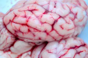
When you pick up a brain model, the folded surface is the cerebral cortex. If a model is made in removable pieces, the cerebrum is usually the part that lifts away first, leaving the deeper structures below. Treat this as a habit of orientation: identify the wrinkly outer layer, then ask what it covers.

Lobes and the link between hemispheres
Each cerebral hemisphere has traditionally been divided into four lobes, per the Queensland Brain Institute. The frontal lobe is the one most often singled out, and the Dana Foundation links it to executive function, with a hand in reasoning and decision-making. For a lobe-by-lobe tour, use the guide to the lobes of the brain.
The hemispheres do not work in isolation. The corpus callosum is a bridge of wide, flat neural fibres that helps relay signals between them, according to the Dana Foundation. On a model cut down the midline, it is the structure that spans the gap between the two cut halves, so it is a good landmark to find early. The page on corpus callosum anatomy covers it in more depth. Other structures sit deeper again, and the guides to basal ganglia anatomy and hippocampus anatomy are the natural next step when you reach those parts of a sectioned model.
Midbrain
The midbrain is the top part of the brainstem, which connects the brain to the spinal cord, as the Queensland Brain Institute puts it. That description gives you both its position and its role. It is a link, not a large mass of tissue in its own right, and it marks where the brain begins to narrow toward the cord.
Hindbrain: medulla, pons and cerebellum
The hindbrain contains the rest of the brainstem, made up of the medulla oblongata and the pons, and also the cerebellum. The cerebellum is a small ball of dense brain tissue nestled right against the back of the brainstem. In functional terms, the Dana Foundation says it handles balance and coordination of movement.
Spatially, that gives a dependable rule for models: find the brainstem first, then look directly behind it for the cerebellum. The brainstem as a whole is responsible for autonomic processes like breathing and heart rate, and it also helps conduct information from the brain to the PNS. The guide to the midbrain pons medulla oblongata sequence goes through each level, and cerebellum anatomy has its own page.
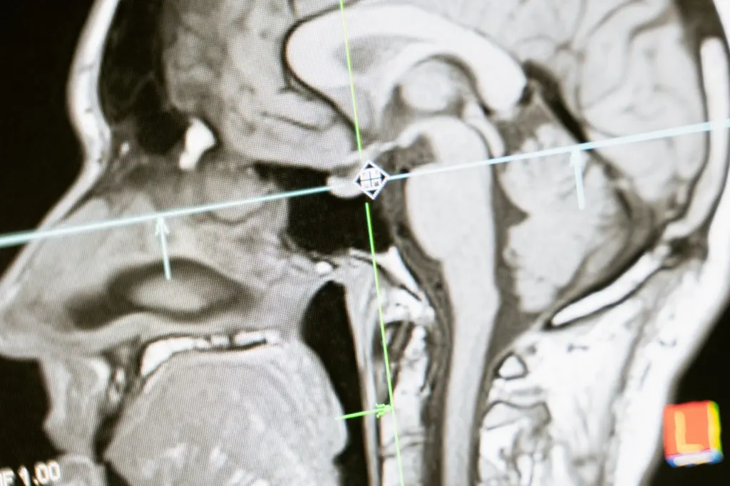
A midline view like the one above is the best single image for checking your mental map. If you can point to the corpus callosum, the brainstem and the cerebellum on it without looking at labels, the regional layout has settled.
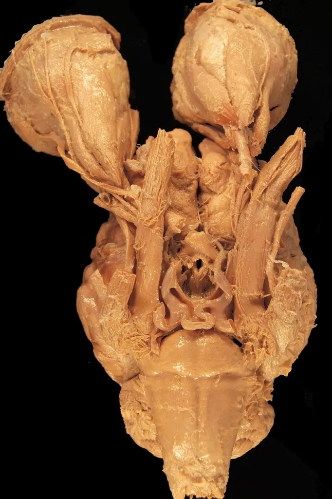
Protection around the brain and cord
The brain is protected by the skull, and the spinal cord is protected by the bony vertebrae of the backbone, according to the Queensland Brain Institute. Both are also protected by three layers of membranes known as meninges. This is why most anatomy models show the bone and membrane as removable layers: the nervous tissue is the focus, but the coverings explain how it is shielded.
For the membranes in detail, see the guide to the meninges layers. Because the same three-layer arrangement applies to the brain and the cord, learning it once pays off in two places.
Blood supply
Blood supply is a separate topic from the nervous tissue itself, but the two are tied together. The Queensland Brain Institute notes that because of the abundant blood supply of grey matter, it is actually more pink-coloured than grey. This is a useful reminder that coloured models are a convention rather than a photograph of living tissue. When you are ready to follow the vessels, start with the blood supply of the brain, then move to the circle of willis arteries, which is the part of the arterial layout that most models highlight.
The covering layers end where the brainstem meets the spinal cord, which makes the cord the next structure to follow.
The spinal cord and its link to the brain
The brainstem connects the brain to the spinal cord, and the spinal cord is part of the CNS together with the brain. That chain answers the question of how the cord connects to the brain: the midbrain is the top of the brainstem, the medulla oblongata and pons make up the rest of it, and the cord continues from there. The brainstem also helps conduct information from the brain to the PNS, so it is the junction between the CNS and the nerves that reach the body.
On a model, trace the column from the cerebrum down through the brainstem and into the cord. If you can name each structure as your finger passes it, you have the connection in working order.
Inside the cord
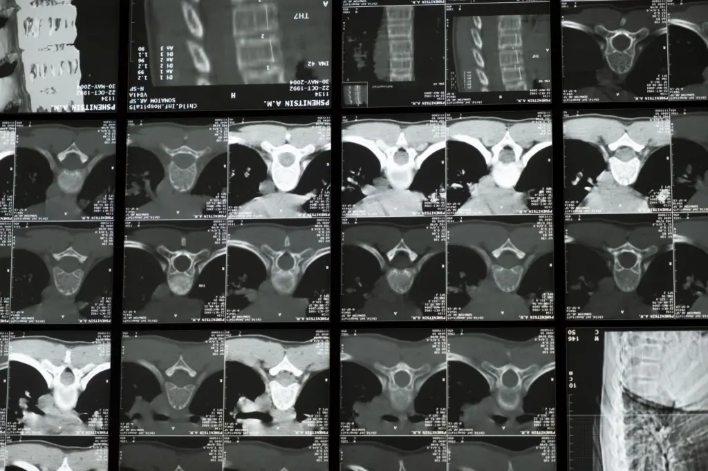
In the spinal cord, grey matter forms the core butterfly shape, as the Queensland Brain Institute describes it. A cross-section model makes this shape obvious, and it is a classic identification point. For the full structure and how it is laid out, use the page on spinal cord anatomy.
From cord to peripheral nerves
Beyond the cord, the PNS takes over. Its nerves and supporting cells branch throughout the rest of the body, so the study path turns outward at this point. The guides to spinal nerves anatomy and cranial nerves anatomy deal with the nerves themselves. For a worked example of a nerve network, brachial plexus anatomy is a good case, and dermatomes and myotomes show how the body is mapped back to the cord.
Whether you study the cord or the nerves, the cell is the common unit, and that is where the finer scale begins.
Neurons and nervous tissue
Neurons are the cells that carry signals, and the Franklin Institute says there are approximately 86 billion of them in the brain. It also states that neurons have dendrites, which receive signals, and axons, which send signals. That receiving-and-sending split is the simplest working description of a neuron, and it is the one to anchor on before you move to cell types. The page on neuron structure develops the detail.
Myelin and white matter
Myelin insulates axons, and the Franklin Institute explains that this allows the signal to travel faster and farther. The Dana Foundation connects this to tissue colour: white matter is made up of the neuron’s axons, sheathed in myelin. That makes white matter the wiring of the nervous system, in contrast to grey matter, which is where the cortex and the butterfly core of the cord are found.
The link between cell structure and tissue type is worth learning together, because it explains why models use two colours. The gray matter vs white matter guide sets the two side by side.
Using models and scans together
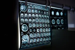
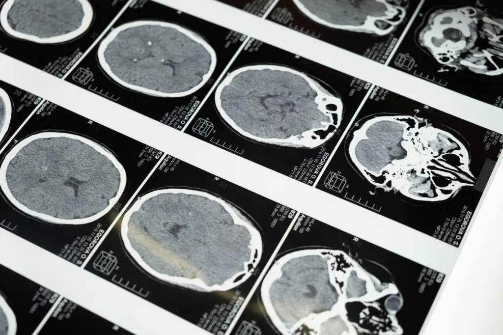
A model gives you the whole structure and its spatial relationships, while a scan shows you a flat slice through a real head. Using both is the fastest way to turn a memorised list into a working map. A model suits you when you need to rotate the structure and see what sits in front of, behind and beneath what. A scan suits you when you need to recognise anatomy in the form it takes in practice.
A routine that works
- Orient before you label. Decide which way the model is facing. Once you know front from back, rules such as the cerebellum sitting against the back of the brainstem become usable instead of abstract.
- Move from outside to inside. Name the cerebral cortex and lobes first, then the corpus callosum, then the brainstem levels, then the cerebellum. That mirrors the way many models come apart.
- Follow the column. Trace from the midbrain through the pons and medulla oblongata and into the spinal cord, saying each name aloud as you pass it.
- Return to the divisions. End each session by sorting what you named into CNS and PNS. This keeps the big categories alive while the details pile up.
- Check a scan. Find the same landmarks on a midline slice, then on a slice cut across the head, and notice how the same structure changes appearance with the cut.
If a model and a textbook seem to disagree, trust the description tied to the structure’s position, such as the top of the brainstem or the back of the brainstem, rather than a colour. Colours are a convention that differs between manufacturers.
Where to go next
After this overview, the right next page depends on the gap you want to close. Use the choices below as a short decision list.
- If cortex areas are the gap, go to the guide to the lobes of the brain, then to the full list of regions on the parts of the brain page.
- If deep structures are the gap, choose basal ganglia anatomy, hippocampus anatomy or corpus callosum anatomy, depending on which structure your model shows.
- If the brainstem and cerebellum are the gap, use the pages on the brainstem levels and cerebellum anatomy.
- If the body is the gap, move to spinal nerves, cranial nerves and the dermatome map.
- If protection and supply are the gap, read the meninges and blood supply pages.
The table below collects the main regions, their position and what each is known for. Use it as a quick check before you open a detailed page.
Region overview
| Region | Where to find it | What it is known for | Planning use |
|---|---|---|---|
| Forebrain (cerebral cortex) | Largest region; the wrinkly outermost layer | Sensory perception, information processing, memory, learning and decision-making | Start here for lobes and cortex areas |
| Midbrain | Top part of the brainstem | Connects the brain to the spinal cord | Use it as the entry point to the brainstem |
| Medulla oblongata and pons | Rest of the brainstem, in the hindbrain | Brainstem as a whole handles breathing and heart rate | Study with the midbrain as a single sequence |
| Cerebellum | Small, dense ball against the back of the brainstem | Balance and coordination of movement | Find the brainstem first, then look behind it |
| Spinal cord | Continues from the brainstem inside the vertebrae | Part of the CNS; grey matter forms a butterfly-shaped core | Pair with spinal nerves when moving to the PNS |
Region descriptions come from the Queensland Brain Institute and the Dana Foundation.
Frequently asked questions
What does the frontal lobe do?
The Dana Foundation describes the frontal lobe as responsible for executive function, with a hand in reasoning and decision-making.
Is grey matter actually grey?
In living tissue it is not. Its dense blood supply gives grey matter a pinkish cast, as the Queensland Brain Institute.
How many neurons does the human brain contain?
The Franklin Institute gives approximately 86 billion neurons in the brain.
How many neurotransmitters are known?
The Franklin Institute reports over 100 identified types of neurotransmitters, the chemical messengers that neurons use to signal across connections (source).