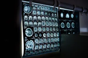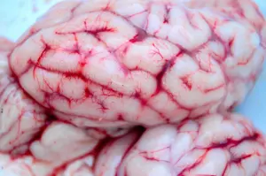

Gray matter vs white matter comes down to composition: gray matter holds nerve cell bodies, while white matter is made of myelinated axons that link them. In the brain gray matter forms a thin surface layer; in the spinal cord the arrangement reverses.
Both tissues belong to one system, and neither makes sense alone. For wider context on how they fit into the organ as a whole, see our guide to brain anatomy.
Gray matter and white matter side by side
Choose gray matter as your focus when the question is where neurons sit and process signals. Choose white matter when the question is how distant regions talk to each other. The table sets the two tissues against the same criteria.
| Criterion | Gray matter | White matter | Planning use |
|---|---|---|---|
| Main components | Nerve cell bodies (soma) holding the nuclei | Myelinated axons extending from the soma | Ask whether you are studying the cells themselves or their cables. |
| What gives the look | Cell-body-rich tissue | Myelin, the fatty coating of axons, makes it white | Colour in a fresh specimen reflects myelin content. |
| Position in the brain | Thin layer on the brain’s surface | Bundles or tracts connecting brain cells to other brain cells | Look to the surface for cell bodies and below it for connecting pathways. |
| Position in the spinal cord | Central core | Wraps around the central core | Expect the brain’s pattern to be reversed in a cord cross section. |
These contrasts follow the descriptions from the Max Planck Society and a Multiple Sclerosis News Today explainer.
What each tissue is made of
The difference between gray and white matter is a difference in cellular parts. Gray matter is where the cell bodies live, and white matter is made of the long fibers that leave them.
Gray matter: cell bodies, synapses and the cortex
Gray matter is composed of nerve cell bodies (soma) that hold the nuclei, according to Multiple Sclerosis News Today. The cerebral cortex is the largest gray matter structure. An Eyewire explainer notes that cortex contains somas and almost all of the brain’s synapses. It also puts the cortex at roughly 50% of the brain’s volume, which makes it a thick layer despite the “thin layer on the surface” description. Treat those two statements as complementary: the sheet is thin in any one place but covers a large area, and it adds up to a large share of the organ.
This is why gray matter is the better entry point for questions about where processing happens. If you want to see how a single cell is built before thinking about tissue, start with neuron structure, then return here to see how those cells are grouped.
White matter: myelinated axons and tracts
White matter includes the billions of myelinated axons extending from the soma. These axons connect brain cells to other brain cells through bundles or tracts. The same Eyewire source describes the fibers as connecting various regions of cortex together. White matter therefore contains the wiring between gray matter regions rather than the cell bodies at their ends.
The coating itself is made by cells called oligodendrocytes, and it speeds up the transmission of information through the brain. That speed is the functional reason white matter exists as a separate tissue: long-distance connections need fast conduction.
Why they look different in a fresh brain
The two tissues look different in a fresh brain because of myelin. The Max Planck Society describes myelin as the fatty coating of nerve cell axons, and states plainly that myelin is what makes the white matter white. Gray matter, built around cell bodies instead of long myelinated fibers, lacks that pale, fat-rich appearance.
In practice, the colour boundary on a brain slice is a boundary between two tissue designs. The darker rim of cortex marks where cell bodies and synapses sit, and the paler region beneath marks where axon bundles pass. Reading a slice this way is more reliable than memorising a list of regions, because it tells you what kind of structure you are looking at before you name it.
Where each is found in the brain and spinal cord
In the brain, gray matter forms a thin layer on the surface, and white matter lies as connecting tracts beneath and between cortical regions. In the spinal cord, the pattern is turned inside out. According to Multiple Sclerosis News Today, white matter wraps around a central core of gray matter.
Brain: surface cell bodies, connecting fibers below
The cortex at the surface carries the cell bodies and the great majority of synapses. The axon fibers that link its regions run in white matter, so a cut through a hemisphere shows a gray rim enclosing paler tissue. Some gray matter structures also sit deeper, among the white matter, which is why the internal capsule discussed below is described relative to neighbouring nuclei.
Spinal cord: gray core, white surround
In a spinal cord cross section, the gray matter forms a central core and white matter surrounds it. If you are comparing a brain slice with a cord section, expect the colours to swap positions: pale tissue outside and darker tissue inside in the cord, darker outside and paler inside in the brain.
White matter fiber types, with the internal capsule as the worked example
White matter fibers are commonly sorted by what they connect: association, commissural and projection fibers. This classification is more useful than learning every tract by name, because it tells you the logic of a pathway at a glance.
Association fibers
Association fibers link regions of cortex to one another within the same hemisphere. This fits the description of white matter as billions of axon fibers connecting various regions of cortex together. If a question is about how two cortical areas on the same side cooperate, association fibers are the relevant class.
Commissural fibers
Commissural fibers cross the midline to connect the two hemispheres, and the corpus callosum is the large bundle that illustrates the class. A midsagittal view shows it as a pale arch running between the hemispheres. Use it as your reference whenever a pathway is described as linking left and right.
Projection fibers and the internal capsule
Projection fibers run between the cortex and deeper structures. The internal capsule is the standard worked example: a projection pathway lying between the thalamus and the lentiform nucleus, seen clearly on a horizontal brain section. Here a white matter band is bounded by gray matter structures on either side, which makes the capsule a compact illustration of how the two tissues sit together.
Because fibers of this class funnel through a restricted space, the internal capsule is a useful landmark when you need to orient yourself on a section. Find the thalamus and the lentiform nucleus first, and the white matter between them is the capsule.

Which tissue to focus on
Your goal decides which tissue deserves attention first. The points below turn the comparison into study choices.
- Studying cells and synapses: start with gray matter, since cell bodies and almost all synapses are found in the cortex. A structure such as the one in our hippocampus anatomy guide is best approached from this cell-level view.
- Studying connectivity: start with white matter and sort pathways by association, commissural or projection class before memorising names.
- Reading a slice: use colour as the first clue, since myelin makes white matter white, then check position against the brain or spinal cord pattern.
- Comparing brain with cord: remember that the cord reverses the arrangement, with gray matter at the centre and white matter wrapped around it.
Frequently asked questions
Which cells make myelin?
Oligodendrocytes produce the myelin that coats nerve fibers, according to Multiple Sclerosis News Today.
Is white matter fully myelinated in adulthood?
Not everywhere. The Max Planck Society notes that the human frontal cortex is not fully myelinated until the fourth decade of life, so myelination continues well past childhood in that region. See the Max Planck Society overview.
What is a tract?
A tract is a bundle of myelinated axons. Tracts are how white matter connects brain cells to other brain cells.