Here is how to memorize cranial nerves for a practical: learn the order with a mnemonic, add a second mnemonic for sensory, motor or both, then rebuild the list from a blank numbered page and attach skull openings last.
The methods below are ranked from the cheapest first step to the most detailed layer, so you can stop when your course stops asking for more. Each one fits inside a wider routine, which is laid out in how to study anatomy.
The twelve nerves at a glance
Humans have twelve pairs of cranial nerves, numbered with Roman numerals I–XII, according to a human anatomy laboratory manual. The numbers follow each nerve’s position on the brainstem, from superior to inferior and then from medial to lateral, so the numerical order is already a memory aid. Learn the table below as your master list, because every method that follows is a way of filling it in from memory.
| Number | Nerve | Sensory, motor or both | Planning use |
|---|---|---|---|
| I | Olfactory | Sensory | Smell. Exits through the cribriform plate of the ethmoid bone. |
| II | Optic | Sensory | Pairs with olfactory as the first sensory-only pair in the functional mnemonic. |
| III | Oculomotor | Motor | Starts the motor-only run of III and IV. |
| IV | Trochlear | Motor | Second motor-only nerve in that run. |
| V | Trigeminal | Both | Branches use different openings, for example V2 through the foramen rotundum, so learn it branch by branch. |
| VI | Abducens | Motor | Motor only, so it sits between two mixed nerves in the pattern. |
| VII | Facial | Both | Internal acoustic meatus and stylomastoid foramen. Carries taste from the anterior two-thirds of the tongue. |
| VIII | Vestibulocochlear | Sensory | The last sensory-only nerve before the mixed run begins again. |
| IX | Glossopharyngeal | Both | First of two consecutive mixed nerves. |
| X | Vagus | Both | Jugular foramen. Motor, sensory and special sensory fibres. |
| XI | Accessory | Motor | Motor only, after the mixed pair IX and X. |
| XII | Hypoglossal | Motor | Hypoglossal canal. Supplies the muscles of the tongue. |
Numbering and openings come from the LibreTexts anatomy lab manual, and the sensory, motor or both labels follow the Integris neurology mnemonic.
Name-order mnemonic: the first method to learn
A name-order mnemonic ranks first because it gives you the one thing every other method depends on, which is the sequence of names. The widely taught version assigns one word per nerve, starting Old (Olfactory), Operators (Optic), Occasionally (Oculomotor) and Troubleshoot (Trochlear), as set out in a nursing study guide. The initials carry on through all twelve nerves.
This method suits anyone who needs the list quickly, including students facing a first lab practical. Its limit is that it stores names and order only. It tells you nothing about what a nerve does, so treat it as scaffolding rather than a finished study plan.
There is no single best mnemonic for cranial nerves. The best choice is the one whose words you can recall without effort, and the strongest approach uses two of them: one for names and one for function.
Functional mnemonic for sensory, motor or both
A second mnemonic covers the classification that name-order phrases leave out. Each cranial nerve is sensory, motor or both, and the phrase “Some Say Marry Money But My Brother Says Big Brains Matter More” encodes that pattern, as Integris Neuroscience describes. Read the first letter of each word in nerve order: S for sensory, M for motor, B for both.
Decoded, the phrase gives the third column of the table above. Olfactory and optic are sensory, oculomotor and trochlear are motor, trigeminal is both, abducens is motor, facial is both, vestibulocochlear is sensory, glossopharyngeal and vagus are both, and accessory and hypoglossal are motor. Say the two phrases side by side, name word then function letter, until the pairs come out together.
To remember what each nerve does, link the class to a concrete detail rather than memorizing the letter alone. Olfactory is sensory because it carries smell. Hypoglossal is motor because it supplies tongue muscles. Vagus is classed as both because it carries motor, sensory and special sensory fibres.
The limit of this method is that “both” hides detail. If your course asks about fibre types, you will need the finer scheme described in the FAQ below.
Blank numbered-list recall
Blank-page recall is the method that tests whether the mnemonics actually worked. The approach described by Integris Neuroscience is to take a sheet of paper, number the lines through to twelve, write out the mnemonic letters, and then fill in each nerve name and its function type.
Do this from memory before you look at the table, then correct it against the table in a different colour. The wrong answers show you which nerves need more work, and those are the rows to revisit next time. This suits students who already know the phrases but feel unsure under exam conditions. It suits anyone short of time less well, because it only reveals gaps and does not teach new material.
Drawing the nerves
Drawing a visual representation of the cranial nerves is a quick option for visual learners, according to Integris Neuroscience. Its value is that it forces you to place each nerve on the brain rather than in a list.
Give the sketch a structure based on where the nerves attach. Two nerves, the olfactory and optic, emerge from the cerebrum or forebrain, and the remaining ten pairs arise from the brainstem, as the LibreTexts lab manual explains. Then add landmarks on the brainstem: the trochlear nerve comes from the posterior midbrain, the trigeminal from the pons, and the glossopharyngeal, vagus and accessory nerves from behind the olive, according to TeachMeAnatomy.
Drawing takes longer than a mnemonic and gives no benefit if you copy the diagram without labelling from memory. Use it after the name order is secure, and redraw it blank at least once. The method pairs well with a regional view of the head, a trade-off covered in regional vs systemic anatomy.
Foramen-based learning
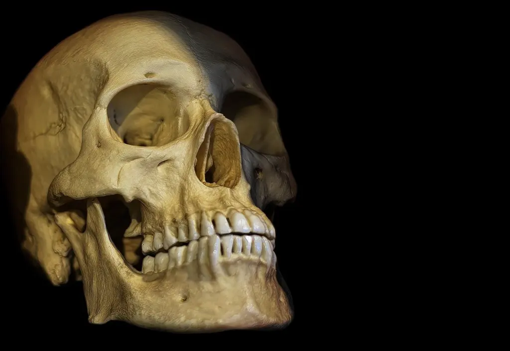
Foramen-based learning ranks last because it is the most detailed layer, but it is the only method that answers the question of where each nerve leaves the skull. Start only when names and classes are stable, and use a real skull, a model or an atlas rather than text alone.
Some openings can be attached to what you already know:
- Olfactory nerve: passes through the olfactory foramina in the cribriform plate of the ethmoid bone, and mediates smell.
- Canal shortcut: a memory tip from TeachMeAnatomy says the nerves with the number two in them, optic and hypoglossal, exit through a canal.
- Trigeminal nerve: each branch has its own opening. The maxillary branch (V2) uses the foramen rotundum and the infraorbital foramen.
- Facial nerve: uses the internal acoustic meatus and the stylomastoid foramen.
- Vagus nerve: leaves through the jugular foramen.
- Hypoglossal nerve: uses the hypoglossal canal and is motor to the muscles of the tongue.
These pairings come from the LibreTexts lab manual. Work through one nerve at a time, name the opening aloud while pointing to it, and add the next nerve only when the previous one is automatic. Check your own course’s list of openings, since the table above gives only the ones the cited manual states.
Decision checklist
Choose the methods in this order, and stop at the layer your course demands.
- Learn the name-order mnemonic until you can recite the twelve names without hesitation.
- Add the functional mnemonic and say each name with its S, M or B.
- Write a blank numbered list from memory and mark the rows you missed.
- Draw the underside of the brain, placing two nerves on the forebrain and ten on the brainstem.
- Attach skull openings one nerve at a time, starting with olfactory, vagus and hypoglossal.
If your exam is mostly written, spend more time on the written-recall steps. If it is a practical on specimens or models, give the drawing and foramen steps more time. Once this routine works for the head, the same layered approach carries over to how to memorize the brachial plexus.
Frequently asked questions
Which cranial nerve has the longest pathway?
The vagus nerve. According to a neurology clinic overview, it extends from the medulla all the way into the abdomen.
Is the trigeminal nerve sensory or motor?
Both. The same overview describes the trigeminal nerve as the largest cranial nerve, with sensory and motor functions.
Why do some textbooks list more categories than sensory, motor and both?
Finer schemes divide cranial nerve fibres into seven modalities. TeachMeAnatomy notes that three of them, SSS, SVS and SVM, are unique to cranial nerves. Learn the simple three-way split first and add the modalities only if your course requires them.