

A 3D human organs model is best chosen by coverage. Pick a systems-based viewer for separable whole-body study, a regional model for abdominal detail, and a scan-based set when the origin of the organ matters.
This guide compares the main kinds of organ set and how they behave. For the wider field beyond organs, start with our overview of each 3D human anatomy model type.
How the organ sets are ordered
The sets below are not ranked from best to worst, because they answer different needs. They run from the widest coverage to the narrowest and most specialised. Four criteria decide which one suits you.
- Organ coverage. A full-body set groups organs by system, while a regional set concentrates on one cavity and names its organs in detail.
- Separability. Some viewers let you rotate, isolate and hide structures. A fixed scene only lets you look from different angles.
- Torso or single organ. A torso set shows how organs sit relative to each other. An individual organ is easier to study closely.
- Scan origin. Some models state that they come from scanned specimens, and others are sculpted. This matters if you want real tissue proportions rather than an idealised shape.
Organ model sets compared
TeachMeAnatomy system models: widest coverage, most separable
TeachMeAnatomy offers interactive 3D models organised by functional physiological system. The groups include the respiratory, gastrointestinal, endocrine, urinary and reproductive systems.
It also handles the question of hiding organs well. Each system model can be rotated, isolated and examined at multiple levels of detail. Supporting structures can be toggled on for context, or switched off so you focus only on the key organs and conduits.
It suits learners who want to follow a system from end to end, for example the respiratory organs including the lungs. The limit is that each model is a system, not a single downloadable object, so it fits study better than reuse in your own project.
Voka abdominal organ model: best for the abdomen
Voka’s abdominal organ model names the stomach, intestines, liver, pancreas, spleen, kidneys and adrenal glands. Choose it when you want the digestive and filtering organs of the abdomen in one scene instead of scattered across systems.

It is a regional model, so it will not show the chest organs. That keeps the view focused, but you will need another set for the lungs and heart. It works well alongside a systems viewer: use Voka to see how the abdominal organs sit together, then switch to a system model to follow one organ group in isolation.
SGU MedArt collection on Sketchfab: best when scan origin matters
The SGU MedArt human anatomy collection states how individual models were made. One entry is titled Human Right Lung Anatomy: 3D Cadaver Scan. The collection also includes a Human Plastinated Heart built by photogrammetry and another labelled as a 3D scan of a plastinated heart. The collection ranges from wet or dry specimens to sculpted models, so check each entry’s title before assuming a model is scan-based.
This is the strongest answer to whether organ models come from real scan data: some do, and the better ones say so in the title. The trade-off is that these are individual organs, not a connected torso. You get close, true-to-specimen detail on a single lung or heart, but you lose the neighbouring structures that a torso model shows.
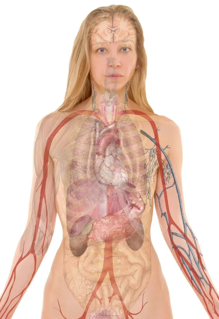
If the heart is your main interest, our guide to 3d heart anatomy goes deeper than a general organ set can.
SciePro commercial library: best for licensed or printed use
SciePro’s commercial model library includes a comprehensive SP ONE model with over 3600 individual structures. The models are described as ready to license and medically reviewed, for animation, education and XR. SciePro also states that, through its collaboration with Stratasys, the models have been tested and validated for professional-grade 3D printing workflows.
This suits studios, publishers and educators who need clear usage rights or a printable model. It is not the right pick for casual study. The sheer number of structures is more than most learners need, and commercial licensing is a cost and a process that a free viewer avoids.
Organ coverage at a glance
| Organ group | How it is presented | Planning use |
|---|---|---|
| Respiratory system | System group in the interactive models | Use it to study the lungs as part of a connected system, with supporting structures toggled on or off. |
| Gastrointestinal system | System group in the interactive models | Use it to follow the digestive organs as a system rather than region by region. |
| Endocrine system | System group in the interactive models | Use it when you want hormone-related organs separated from the rest of the torso. |
| Urinary system | System group in the interactive models | Use it to isolate the urinary organs, then compare with the kidney in an abdominal scene. |
| Reproductive system | System group in the interactive models | Use it when this system needs to be viewed separately from the abdominal organs. |
| Abdominal region | Named list: stomach, intestines, liver, pancreas, spleen, kidneys, adrenal glands | Use it to see how neighbouring abdominal organs sit relative to each other. |
The system groups come from TeachMeAnatomy’s interactive system models, and the abdominal list from Voka’s abdominal organ model.
Choosing between viewing, downloading and printing
Viewing online
For study, an online interactive viewer is the simplest route. TeachMeAnatomy’s system models and the SGU MedArt collection are both published as online collections you can open and explore. Use them when you only need to look, rotate and isolate organs, and you do not need the file itself.
Hiding and separating organs
Separability is the feature that most changes how useful a set is. Systems-based viewers that let you isolate a system and toggle supporting structures serve students well. A fixed single-organ scene is better for close inspection of one organ than for understanding how organs relate. If you want to see where an organ sits, choose a torso or regional model and not an isolated scan.
Downloading and file format
Match the file to your purpose, not to a favourite format. If you only view organs, you need no download. If you plan to print, look for a vendor that states its files are validated for printing, as SciePro does with Stratasys. Check the format listed on each download page before you commit, and confirm the licence covers your use.
Putting a set to work
A practical order is to learn the system view first, then look at one region, then examine a scanned single organ. Muscle layers sit outside the organ sets, so for that layer see our guide to 3d muscle anatomy.
Frequently asked questions
What do the liver and kidneys do in an abdominal organ model?
Voka’s abdominal model describes the liver as performing detoxification, protein synthesis and production of biochemicals necessary for digestion. It describes the kidneys as filtering blood to produce urine.
Can I embed an organ viewer on my own platform?
For Voka’s abdominal model, embedding on a partner platform is arranged by getting a quote, so it is not a simple copy-and-paste option.
Are free organ models easy to find?
Free options exist, but one developer discussion on the three.js forum observes that the real good stuff is paid. If you need models you can redistribute, expect to look at licensed libraries.