A sudden fright and a quiet meal call on the sympathetic vs parasympathetic nervous system in opposite ways: the sympathetic division prepares the body for action, the parasympathetic restores baseline, and both are always active to some degree.
For the wider structure of the central and peripheral nervous system, start with brain anatomy. For how the brainstem and other regions fit into the picture, see parts of the brain and their functions.
Criteria for comparing the two divisions
The autonomic nervous system controls involuntary body functions, and Lifestack describes it as having two primary divisions, the sympathetic (SNS) and the parasympathetic (PNS). The comparison is clearest when each division is judged on the same short list of criteria, in this order.
- Overall job. Decide whether the division mobilises the body for action or returns it to baseline. This is the single most useful distinction and the one to learn first.
- Outflow from the central nervous system. Note where the fibres leave the brainstem or spinal cord, because the names thoracolumbar and craniosacral come from this.
- Ganglion position. Check whether the relay station lies near the spinal column or close to the target organ.
- Transmitter at the target. Identify whether the final neuron releases noradrenaline or acetylcholine.
- Effect on the organ. Finish with the heart, pupils and gut, where the two divisions pull in opposite directions.
Working through the list in this order stops the common mistake of memorising organ effects without knowing why they occur. The table below condenses the criteria that can be stated as direct contrasts.
Side-by-side comparison
| Criterion | Sympathetic | Parasympathetic | Planning use |
|---|---|---|---|
| Overall role | Prepares the body for action | Restores the body to baseline | Use as the anchor for every other row |
| Transmitter at the target organ | Norepinephrine (noradrenaline) | Acetylcholine | Memorise as a pair; the labels differ in British and US texts |
| Heart rate | Increases, pumping more blood to muscles | Slows toward the resting baseline | Treat as the standard opposing-effect example |
| Pupils | Dilate to take in more light and visual information | Constrict | Best single organ for testing recall |
| Digestion | Slows, as energy is redirected from non-urgent processes | Resumes, with digestive enzyme production increasing | Explains why a stressed person may lose appetite |
These contrasts follow the comparison set out by Lifestack, which presents the two divisions as opposing partners.
Where the fibres leave the central nervous system
The two divisions are separated most formally by their outflow. Sympathetic fibres leave the spinal cord and are called thoracolumbar. Parasympathetic fibres leave the brainstem and the lower end of the spinal cord and are called craniosacral. Learning the pair of names is the quickest route to remembering the layout, because each name describes the region of origin.
Thoracolumbar sympathetic outflow
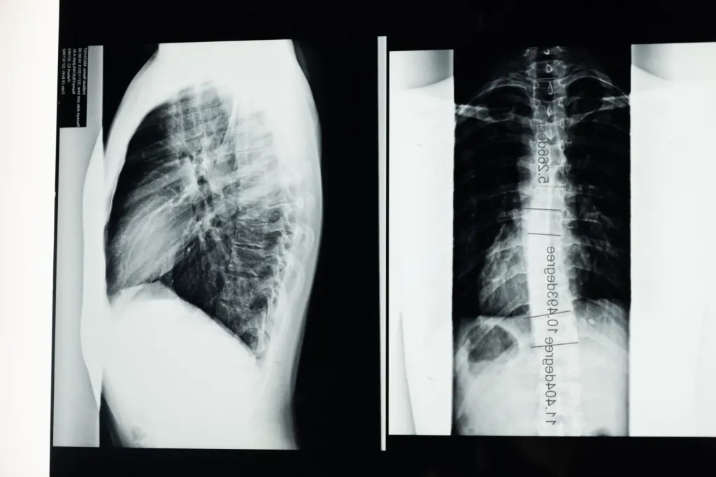
The sympathetic division is tied to the trunk, which is why vertebral landmarks matter when studying it. Imaging of the thoracic spine gives a useful orientation to the region this outflow occupies, though a radiograph shows bone and not the nerve tissue itself.
Craniosacral parasympathetic outflow
The best-known parasympathetic pathway is the vagus nerve. Lifestack calls it the primary driver of parasympathetic activation and the longest cranial nerve in the body, running from the brainstem through the chest and into the abdomen. That course explains why one nerve can slow the heart and also restart digestion. The sacral part of the outflow serves pelvic organs and is the second half of the craniosacral label.
Ganglion position and a short ganglia list
Autonomic pathways use two neurons in sequence, and the relay between them is a ganglion. The position of that relay is a reliable way to tell the divisions apart. Sympathetic ganglia lie close to the vertebral column, while parasympathetic ganglia lie near or within the organ they serve.
- Sympathetic chain ganglia. These sit in a paravertebral chain beside the spine and give the sympathetic division its wide, body-wide reach.
- Ciliary ganglion. This parasympathetic ganglion lies in the orbit and is part of the pathway to the pupil, so it links ganglion position to a visible organ effect.
Students who also study segmental supply to skin and muscle can compare this pattern with dermatomes and myotomes, where the spinal level likewise determines what is supplied. A ganglia list is best learned as a pairing of each ganglion with the organ or region it serves, not as a bare set of names.
Neurotransmitters: acetylcholine and noradrenaline
At the target organ, the sympathetic division uses norepinephrine, known in British usage as noradrenaline, and the parasympathetic division uses acetylcholine, according to Lifestack. The sympathetic division also triggers release of cortisol and adrenaline (epinephrine) from the adrenal glands, which extends its effect beyond the nerve endings themselves.
Transmitters at each synapse of the pathway are a standard topic in autonomic teaching; the Pearson anatomy and physiology course lists neurotransmitters of the ANS as its own lesson within the autonomic nervous system chapter. When revising, keep the target-organ pairing separate from the ganglionic step, since mixing them is a frequent source of exam errors.
Organ effects: heart, pupils and gut
The organ effects follow directly from each division’s job. Where the sympathetic division mobilises resources, the parasympathetic division withdraws that mobilisation.
Heart and lungs
Sympathetic activity raises heart rate and pumps more blood to muscles, and bronchioles in the lungs dilate to allow more oxygen intake. Parasympathetic activity slows the heart toward its resting baseline. The heart is therefore the cleanest example of one organ under two opposing influences at once.
Pupils
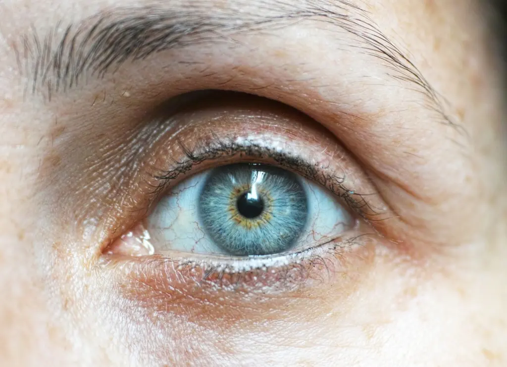
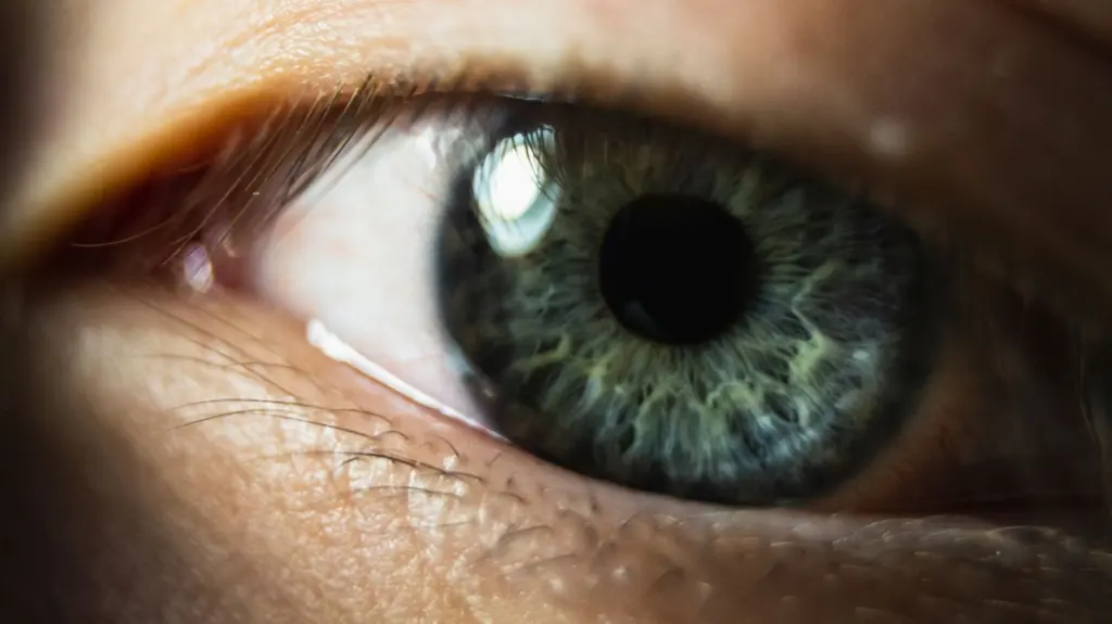
Pupil size shows the opposition at a glance: the sympathetic division dilates the pupils and the parasympathetic division constricts them. A photograph shows the result but not the pathway, so the ciliary ganglion described above supplies the anatomical explanation.
Gut
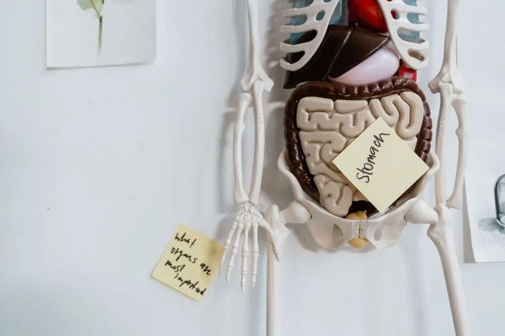
During sympathetic dominance digestion slows, because energy is redirected away from non-urgent processes. When the parasympathetic division takes over, digestion resumes and digestive enzyme production increases. This is why the parasympathetic division is often described through rest and digesting, and why the abdomen is covered by the vagus nerve’s course.
Choosing the right frame for study
Treat the two divisions as balanced partners, not as an on-off pair. Both are active at all times to some degree, so the useful question is which one dominates in a given state. Sleep is the strongest parasympathetic state the body reaches, while an emergency pushes the balance toward the sympathetic side.
- For a quick overview. Learn the overall roles and the heart, pupil and gut examples. These three organs cover the logic of the whole system.
- For anatomy exams. Add outflow level, ganglion position and the transmitter pair, because these are the structural features that separate the divisions.
- For physiology or clinical reasoning. Concentrate on the vagus nerve and on organ effects, since they connect anatomy to observable changes in heart rate, pupil size and digestion.
A common edge case is assuming that a rise in one division means the other has switched off. The supported picture is a shift in balance, not a switch between two states.
Frequently asked questions
| Question | Answer |
|---|---|
| Are the sympathetic and parasympathetic divisions ever active at the same time? | Yes. Both are always active to some degree, and the body’s state depends on which one is dominant. |
| Which nerve drives parasympathetic activation? | The vagus nerve, the longest cranial nerve in the body, running from the brainstem through the chest and into the abdomen. |
| What does the autonomic nervous system control? | It controls involuntary body functions and has two primary divisions, the sympathetic and the parasympathetic. |
| Which state is the strongest parasympathetic state? | Sleep is the most powerful parasympathetic state the body achieves. |