Struggling to see where one region of a brain model ends and the next begins? The four lobes of the brain are the frontal, parietal, temporal and occipital, and grooves called sulci mark where several of them begin and end.
This guide works from landmarks first, so you can trace each lobe on a three-dimensional model before learning its job. For the wider picture, start with our overview of brain anatomy, or see parts of the brain and their functions for structures beyond the cerebral lobes. Structures outside the cerebrum, such as the cerebellum, are covered in cerebellum anatomy.
Problem: you cannot tell which lobe is which
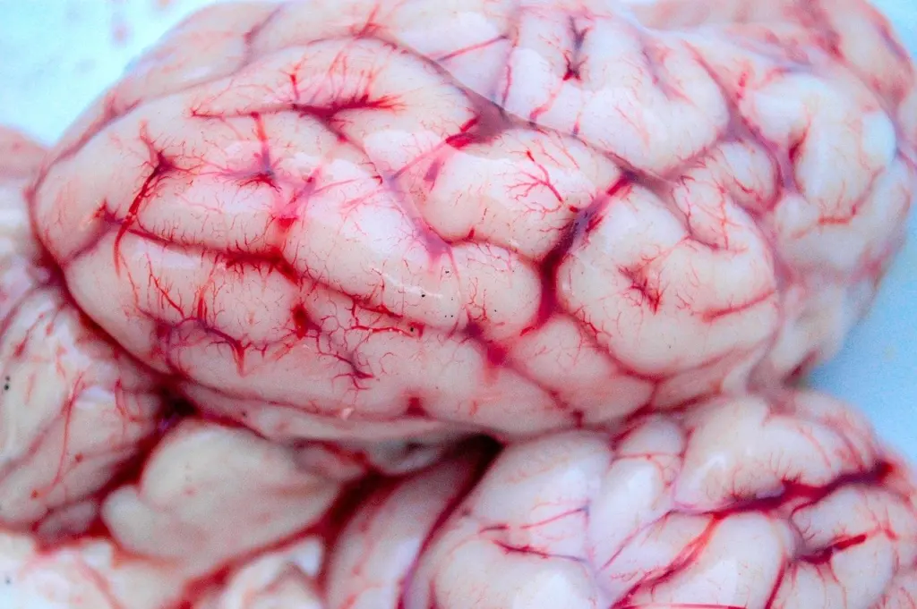
The cerebrum can be separated into four distinct lobes, according to BrainFacts. Yale’s brain and language resource names them as the frontal, parietal, temporal and occipital lobes. Colour-coded diagrams make this look easy, but an uncoloured model or a real specimen gives you nothing to go on except the surface itself.
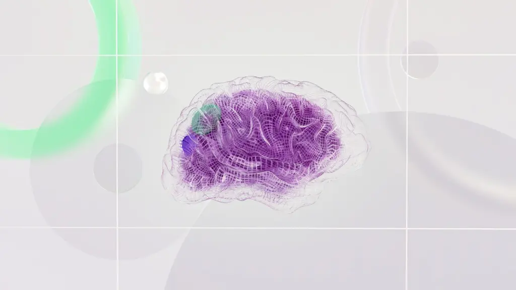
Solution: learn the position, then the borders. Position gives you a first guess. The frontal lobe sits at the front, and the occipital lobe is at the back of the brain, behind the parietal lobe and temporal lobe, as the University of Washington’s neuroscience pages describe it. Sulci then confirm the guess, and the next section shows how.
Solution: trace the boundaries with sulci
A sulcus is a groove in the brain surface, and the gyri are the bumps between the grooves. Two grooves do most of the work in telling the lobes apart: the central sulcus and the lateral sulcus. Learn these two first and the other lobes fall into place around them.
Central sulcus: frontal versus parietal
The central sulcus is the dividing line between the front and the middle-back of the hemisphere. The frontal lobe is located in front of the central sulcus, while the parietal lobe is located behind it. If you can find this groove on a model, you can name the lobe on either side of it without any colour cue.
When the groove is hard to spot, use the parietal lobe’s general position as a cross-check. MedlinePlus places the parietal lobes behind the frontal lobe, towards the back of the head.
Lateral sulcus: the upper edge of the temporal lobe
The lateral sulcus, also called the lateral fissure, is the deep groove that runs along the side of the hemisphere. The temporal lobe is located below it. On a side view, find the groove and then look at the tissue hanging beneath it. That tissue is the temporal lobe.
Occipital lobe: defined by position
The occipital lobe is the simplest to identify because it occupies the posterior end of the hemisphere, behind the parietal and temporal lobes. Once you have named the lobes on either side of the central and lateral sulci, the remaining tissue at the very back is occipital.
Diagnostic table: from clue to lobe
| What you see | Landmark clue | Lobe | Planning use |
|---|---|---|---|
| Tissue ahead of a deep groove running across the top of the hemisphere | In front of the central sulcus | Frontal lobe | Find the central sulcus first, then step forward. |
| Tissue just behind that same groove | Behind the central sulcus | Parietal lobe | Step backward from the central sulcus to confirm. |
| Tissue hanging beneath the long groove on the side of the hemisphere | Below the lateral fissure (lateral sulcus) | Temporal lobe | Use a side view and find the lateral sulcus first. |
| Tissue at the rear of the hemisphere | Behind the parietal and temporal lobes | Occipital lobe | Name the other lobes first, then the rear tissue is occipital. |
Boundary descriptions are drawn from the University of Washington’s lobe reference and the MedlinePlus anatomy video summary.
Decision guide: what each lobe is associated with
Once the borders are clear, function helps you remember which lobe is which. These are the principal functions attributed to each lobe, not an exhaustive map of cortical areas, and they are best used as memory anchors alongside the landmarks above.
- Frontal lobe: choose this for planning and movement. The University of Washington describes it as concerned with reasoning, planning, parts of speech and movement (motor cortex), emotions and problem-solving. MedlinePlus adds that voluntary body movements are controlled by a region of the frontal lobe and that this is where we shape emotional reactions and expressions.
- Parietal lobe: choose this for bodily sensation. It is concerned with perception of stimuli such as touch, pressure, temperature and pain.
- Temporal lobe: choose this for sound and memory. MedlinePlus states that all sounds are processed in the temporal lobe. The University of Washington adds recognition of auditory stimuli and memory, with the hippocampus named in connection with memory.
- Occipital lobe: choose this for vision. It analyzes visual information from the retina, and the University of Washington describes it as concerned with many aspects of vision.
If you are asking what the frontal lobe does, the practical answer is that it covers the broadest mix: thinking, movement and emotional expression. That breadth is why it is worth locating the central sulcus before anything else, since the groove marks its back edge.
Why the surface is folded into gyri and sulci
The cerebral cortex is the outside portion of the cerebrum, also called the gray matter, according to MedlinePlus. In neuroanatomy this folded outer sheet is referred to as the neocortex. Its grooves and folds increase the cerebrum’s surface area.
The University of Washington gives the scale: the total surface area of the cerebral cortex is about 324 square inches. It explains that the folding created by the sulci and gyri increases the amount of cerebral cortex that can fit in the skull. A smooth surface would need far more room, so the folds are what let this much cortex sit inside the head.
For a student, the point is practical. Every groove is a possible landmark, but only a few are used to define the lobes. Treat the central and lateral sulci as your reference grooves, and treat the gyri as the named ridges that sit beside them.
What the cortex is made of
In the brain, the surface of the cerebral cortex is a layer of gray matter, as Michigan State University’s neuroscience text puts it, with white matter lying beneath. Gray matter regions are comprised of cell bodies and dendrites. That is why a cut or stained section of cortex looks different from the tissue below it, and why anatomists talk about the cortex in terms of layers.
This composition is shared by all four lobes. The lobes differ in location and in the functions attributed to them, but each is covered by the same kind of gray matter surface.
Troubleshooting common mix-ups
- The model does not match the textbook picture. Do not assume the model is wrong. Each person has a unique pattern of gyri and sulci, so minor groove patterns vary. Rely on the central and lateral sulci rather than on the exact shape of a bump.
- You cannot tell a sulcus from a gyrus. Remember that sulci are the grooves and gyri are the raised bumps between them. A landmark that you need to trace along is nearly always a groove.
- The parietal and temporal lobes seem to blur together. Go back to the lateral sulcus. Tissue beneath it is temporal. Tissue behind the central sulcus and above the lateral sulcus belongs to the parietal region.
- You are looking at a medial or top view with no lateral groove in sight. Switch to a side view before naming the lobes, because the lateral sulcus is the clearest landmark for the temporal lobe there.
Frequently asked questions
| Question | Answer |
|---|---|
| Is a sulcus the same as a fissure? | The University of Washington uses the terms interchangeably, describing the sulci (or fissures) as the grooves and the gyri as the bumps. The lateral sulcus is also called the lateral fissure. |
| Does everyone’s cortex fold in the same way? | No. Each person has a unique pattern of gyri and sulci, although the main landmark grooves are used to define the lobes. |
| Why does folding matter for brain processing? | BrainFacts says the folding amplifies the brain’s processing power by making more space for synaptic connections. |
| Where are the parietal lobes in relation to the ears? | MedlinePlus describes them as behind the frontal lobe, towards the back of the head and above the ears. |