| Part | What it does | Planning use |
|---|---|---|
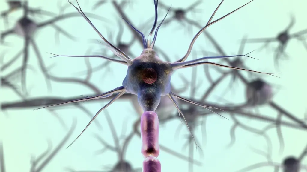
| Dendrites | Tree-like extensions that reach away from the cell body and receive messages at synapses | Trace these first on a diagram to find where a neuron takes in information. |
| Soma (cell body) | Holds the nucleus, smooth and rough endoplasmic reticulum, Golgi apparatus and mitochondria | Use the nucleus as your anchor point when labelling any neuron. |
| Axon hillock | Integrates signals from multiple synapses before the axon | Mark it as the hand-off between receiving and sending. |
| Axon | Tube-like process that propagates the integrated signal to the axon terminals | Follow it away from the soma to find the output end. |
| Myelin sheath and nodes of Ranvier | Insulation that speeds conduction, with gaps where the signal is recharged | Treat myelin as glial insulation around the axon, not as a neuron part. |
| Synapse | The space between the end of an axon and the tip of a dendrite of another neuron | Label it as a junction between two cells. |
Part descriptions follow the NICHD overview of nervous system parts and the Lumen Learning chapter on neurons.
Neuron structure centres on a cell body (soma), tree-like dendrites that receive messages and an axon that carries the signal onward. Myelin, made by glial cells rather than the neuron itself, speeds conduction.
This page treats the neuron as a single cell and sorts neurons by shape and by job. For the larger organs and regions these cells build, see our guide to brain anatomy.
Receiving and integrating: dendrites, soma and axon hillock
A neuron is the basic unit of the nervous system, and its input side is built from dendrites and a cell body. The NICHD describes a neuron as a cell body that includes the nucleus, plus special extensions called axons and dendrites.
Dendrites
Dendrites are tree-like structures that extend away from the cell body to receive messages. The messages arrive at synapses. The scale of that input can be large: the dendrites of a Purkinje cell receive contact from as many as 200,000 other neurons, according to Lumen Learning. A neuron with a heavily branched dendritic tree is therefore built to collect many inputs, which is why dendrites are the first thing to trace on any diagram.
Soma and axon hillock
The soma, or cell body, contains the nucleus, smooth and rough endoplasmic reticulum, the Golgi apparatus and mitochondria. Beyond it sits the axon hillock, a cell body structure that integrates signals from multiple synapses before the axon takes over. For revision, think of the soma and hillock as the place where many incoming messages become one outgoing signal.
Conducting: the axon, myelin sheath and nodes of Ranvier
The axon is a tube-like structure that propagates the integrated signal to specialised endings, the axon terminals. Neurons usually have one or two axons, so the useful rule when reading a diagram is to look for the single long process that leaves the soma and does not branch like a dendrite.
What the myelin sheath does
Myelin is an insulating sheath that wraps the axon and speeds conduction. Many students assume it belongs to the neuron, but Lumen Learning states that the myelin sheath is not actually part of the neuron: glial cells produce it. The NICHD likewise lists the creation of myelin insulation, which helps move nerve impulses, among the roles of glia. The glial cells themselves are outside the scope of this page, which keeps to the neuron.
Nodes of Ranvier
The sheath is not continuous. The gaps between sheathed stretches are called nodes of Ranvier, and they are the sites where the signal is recharged. When you label a myelinated axon, mark the insulated segments and the nodes as alternating features. If you want to see how this insulation shows up when whole tissues are compared, read gray matter vs white matter.
Passing the message on: synapse structure
A synapse is the space between the end of an axon and the tip of a dendrite from another neuron, as defined by the NICHD. That definition has a practical consequence for labelling. A synapse involves two cells, so it belongs to neither neuron alone. The axon terminal on one side and the receiving dendrite on the other are the parts that belong to individual neurons.
The same junction explains the earlier dendrite and hillock facts. Dendrites receive messages at synapses, and the axon hillock sums the signals arriving from many of them. If a diagram shows the end of an axon sitting beside a dendrite, you are looking at the output side of one cell meeting the input side of the next.
Neuron types by number of processes
Neurons are broadly divided into four basic types by the number of processes extending from the soma: unipolar, bipolar, multipolar and pseudounipolar. The table below sets them side by side.
| Type | Processes from the soma | Planning use |
|---|---|---|
| Unipolar | Only one process | Learn the definition so you can tell it apart from pseudounipolar. |
| Bipolar | One axon and one dendrite | Count the two processes at opposite ends of the soma. |
| Multipolar | One axon and multiple dendrites; the most common type | Treat this as the default shape when a diagram is unlabelled. |
| Pseudounipolar | A single process from the soma that later branches into two | Look for the branching point after the single stem. |
Type definitions are from the Lumen Learning chapter on neurons.
Multipolar neurons
Multipolar neurons are the most common type, and each contains one axon with multiple dendrites. A stained multipolar cell on a Digital Histology slide shows the nucleus and Nissl substance in the cell body, with two dendrites and an axon. The axon lacks rough endoplasmic reticulum, which is a helpful cue for telling it apart from the dendrites.
Bipolar and pseudounipolar neurons
The difference between these two comes down to how the processes leave the soma. A bipolar neuron has a separate axon and dendrite. A pseudounipolar neuron has one process that starts like a unipolar cell and then divides. Most sensory neurons are pseudounipolar, with an axon that branches into two extensions, one reaching the dendrites and the other the spinal cord. Choose the multipolar pattern for a cell with many branching dendrites, bipolar for exactly two processes, and pseudounipolar when a single stem splits.
Functional types: sensory, motor and interneurons
The functional scheme asks what a neuron does rather than what it looks like. NICHD states that sensory neurons detect light, sound, odor, taste, pressure and heat and send messages to the brain. Motor neurons transmit messages from the brain to the muscles to generate movement. Interneurons are the third class in this scheme, distinct from sensory input cells and motor output cells.
The two schemes overlap rather than compete. Because most sensory neurons are pseudounipolar, a sensory cell can be described by its job and by its shape at the same time. To place these cells within the whole system, see central vs peripheral nervous system.
Reading a neuron on a histology slide
At histological scale you are looking at one stained cell at a time, so work from the soma outward. A short routine keeps labelling consistent:
- Find the soma. Locate the nucleus, and in a stained multipolar cell look for Nissl substance in the cell body.
- Trace the dendrites. Follow the tree-like extensions that reach away from the cell body.
- Identify the axon. Look for the process that leaves the soma without rough endoplasmic reticulum.
- Mark any insulation. If the axon is sheathed, label the myelin and the nodes of Ranvier between sheathed stretches.
- Note the junction. Label the synapse where an axon end meets another cell’s dendrite.
Common mix-ups
- Myelin as a neuron part. Glial cells produce the sheath, so list it as an insulator around the axon.
- Unipolar versus pseudounipolar. A pseudounipolar cell has a single process from the soma that later branches into two, so check for the branch before deciding.
- Synapse ownership. The synapse is the space between two cells, so avoid assigning it to just one neuron.
- Type versus function. Shape (multipolar, bipolar, pseudounipolar) and job (sensory, motor, interneuron) are separate labels that can both apply to one cell.
Frequently asked questions
How many neurons does the human brain contain?
The NICHD gives about 100 billion neurons for the human brain, describing the neuron as the basic unit of the nervous system.
Can a neuron have more than one axon?
Yes. Neurons usually have one or two axons, according to Lumen Learning, although the typical multipolar neuron has one axon with multiple dendrites.
Are unipolar neurons found in humans?
No. Unipolar neurons are not found in vertebrates. The closest human counterpart is the pseudounipolar sensory neuron, whose single process branches into two.